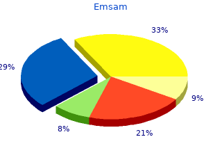
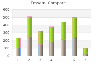
Emsam
2018, Dillard University, Sebastian's review: "Emsam 5 mg. Only $0,83 per pill. Purchase online Emsam no RX.".
If the studies are performed for the purpose of differentiation of intra- cranial toxoplasmosis from lymphoma in the immunosuppressed patient order emsam 5mg with visa anxiety side effects, recent treatment for toxoplasmosis might produce a false positive uptake generic emsam 5mg without prescription anxiety symptoms explained. Reporting The report should include the radiopharmaceutical used, dose, route of injection, waiting period, clinical history and the reason for referring the patient for the study. If sedation has been given it should be mentioned, as well as any adverse reactions. The report should include and mention the findings of other morpho- logical imaging modalities and their correlation with the nuclear medicine procedure. The intensity of uptake should be graded as low, medium or high, together with the size of the lesion as accurately as possible, and its location. Principle The trinity of metabolism–function–blood-flow of the brain are closely interrelated. The subtraction of rest images from activated images enables a clearer identification of activated regions of the brain. Activation paradigms include visual, audio and finger motion stimulations as well as speech and thinking. Activation studies may elucidate higher brain functions in healthy volunteers and neuropsychiatry patients. Clinical indications The indications are the same as those for the cerebral perfusion studies (Section 5. Radiopharmaceuticals Positron emitting radiopharmaceuticals are used for metabolic imaging. Three aspects of cerebral metabolism are of interest clinically, namely glucose and oxygen utilization and protein synthesis. Oxygen-15-O2 can be continuously inhaled, with little dissolved in plasma and most bound to haemoglobin. It is 15 the bound portion of O that is transported to, and utilized by, the brain. Carbon-11-methionine shows protein synthesis and is used mainly for brain tumour imaging. Protocols Patient preparation and pre-test precautions are similar to those described for perfusion. Because of the very short half-life of radionuclides, brain metabolic imaging may be repeated at short intervals to facilitate assessment of different brain states. Acquisition is usually accompanied by a transmission session for attenuation correction. If a kinetics analysis is required, dynamic or fast repeated acquisi- tions are needed. Data processing and interpretation Cerebral metabolic images are similar to those of cerebral perfusion. Usually the metabolic and perfusion images are similar in pattern under normal circumstances. Metabolic images should be interpreted with the structural data available, and co-registration techniques are of great value. Clinical indications Most receptor–transporter studies have been performed to evaluate movement disorders, epilepsy and psychiatric illnesses, but clinical indications are still investigational. Radiopharmaceuticals The radiotracers used in the functional imaging of the brain are listed in Table 5. Protocol Preparation, basic requirements and operational procedures are almost identical to those used in perfusion and metabolic studies. Intervention, for example audiovisual stimulation, task performance tests and complicated conditioning, are more widely used in neuroreceptor studies. Since the receptor study requires detailed spatial and timing information, the use of specific analysis and image fusion with an anatomically informative modality (e. Special notes for receptor imaging (a) Neuroreceptors Neuroreceptors are membrane bound proteins that bind to exogenously administered agents in addition to endogenously released neurotransmitters. There are two types of receptor: (1) Those that are a part of the structure of the so-called ligand-gated channels that directly affect membrane potential and ionic permeability; (2) Those that act by affecting intracellular second messengers via G proteins. They have no pharmacological effects because of the very small amounts administered.
We Introduction/Background: Poliomyelitis was generally consid- report a median and ulnar neuropathy which was not associated ered a non progressive disease and paralytic polio survivors live with chemotherapy and radiaotherapy 5mg emsam free shipping anxiety symptoms of. However purchase emsam 5 mg with amex anxiety symptoms upset stomach, late com- female patient presented with a tingling sensation on right hand, plications may occur. And she had chemotherapy of a 45 year old female patient with prior acute poliomyelitis and radiotherapy. The clinical presentation was a left monople- set, and a few months later tingling sense on right hand was onset. She has a long leg brace but she didn’t want to wear it so In physical examination, there was tingling sense on right hand. She was referred to our unit And circumference of right upper limb was increased by 5~6cm with a chief complaint of easy fatigability of the right arm with more than left side, forearm hardness in median and ulnar nerve paresthesia. There was no reduced muscular strength, and Physical examination revealed an atrophy of the hypothenar emi- tinnel, phalen sign are all positive at right. A protocol of rehabilitation was instituted and we diameter change was observed at carpal tunnel level and absent at encouraged the patient to regularly wear the leg brace and we forearm mid-portion. Conclusion: Neurological a carpal tunnel syndrome, but it is ruled out on the basis of elec- complications mainly consist of the post polio syndrome. The patient was conducted follow up electrophysi- entrapment syndromes of the upper limb are less frequent and ologic study in 2015, its fnding was suitable for right median & can be caused by the use of crutches or wheelchairs. Results: We supposed that neuropathy was pro- be prevented by an appropriate medical follow-up, patient coun- gressive because peripheral nerves were vulnerable due to diabetes seling and suitable measures. Material and Methods: This case is original be- cause of the etiopathogenesis of its neurological damage. Results: Introduction/Background: Focal peripheral neuropathy is one of We report a case of a 27-year-old man. He was a victim of a serious the most common clinical syndromes that are seen in daily routine car accident, which led to a bilateral fracture of the obturator ring, practice of neuromusculoskletal physicians, including surgeons, a fracture of the right sacral ala, a sagittal trans-sacral foraminal rheumatologists, neurologists and physiatrists. Material and Meth- fracture and a fracture of the right transverse processes of L4 and ods: Electrodiagnosis is so far gold standard for diagnosis of fo- L5. Results: Nowa- tion revealed walks with steppage gait with a foot levator muscles days with emergence of imaging techniques, application of these weakness estimated 1/5 and hypoesthesia in the right L5 territory. There is different Cons and Pros spondylolisthesis and a major bone remodeling of the right sacral about these 2 diagnostic tools. Indeed, the scan sonography mandate physiatrists to apply this invaluable diagnos- revealed a compression of the right L5 root in its extra-foraminal tic tool in their daily professional practice. Entrapment syndrome portion due to bone remodeling of the right sacral ala fracture. Con- especially carpal tunnel syndrome is one of the most common clusion: Neurological complications of fractures of the sacrum and clinical issues referred and treated by physiatrist. A care- to understand cons and pros of routine electrodiagnostic medicine ful and a repeated neurological examination is required in order to techniques and evolvling ultrasonographic one for detection of avoid delay in diagnosis. Material and Methods: A total of 17 selected patients with caused by loss of perspiring on the affected side of the face which was pain in the lateral aspect of the elbow, Recorded the medical his- consistent with postganglionic sympatic injury at carotis artery level tory, Tested objective examination, the compound muscle action on left side. In which 3 patients were cial in physical medicine and rehabilitation departments to detect and interosseous nerve lesion and 1 patient was superfcial radial nerve prevent other health problems. The remaining 5 patients were diagnosed with demyelina- chains are vulnerable as well as peripheric nerves and plexus to fre- tion of radial nerve. The diagnosis was confrmed by the medical his- exam is very crucial in physical medicine and rehabilitation depart- tory, objective examination and to compare the latency difference ments to detect and prevent other health problems. Son1 History of trauma or operation 3 (17%) 1Korea University Guro Hospital, Physical Medicine and Rehabili- Posterior lateral pain of elbow joint 12 (71%) tation, Seoul, Republic of Korea Weakness of thumb or fnger extension 3 (17%) Introduction/Background: Recently, ultrasonography has been used Weakness of wrist extension 1 (0. One bullet embedded anterior neck subcutenously and the other 1 1 1 1 1 bullet left from anterior midline neck region. Son tion revealed vocal cord injury, left carotis interna artery wall lac- 1Korea University Guro Hospital, Physical Medicine and Rehabili- eration, left hemothorax and left thyroid gland open wound. Material tation, Seoul, Republic of Korea J Rehabil Med Suppl 55 Poster Abstracts 171 Introduction/Background: Appropriate imaging and electrodiag- effect on the offspring’s development and will increase in depres- nostic studies are essential part of the evaluation of the patient sion- and anxiety-like behavior and alteration in social behaviors.
Head brain with polished edge - the folds are broad and flat and gyri between them were shallow purchase 5 mg emsam free shipping anxiety 12 step groups. By convexity there is a heavy white purulent exudate buy cheap emsam 5 mg on-line anxiety symptoms psychology, located subarachnoidal, filling gyri and spreading on the folds. Organ diagnosis is made in view of the rear surface of the preparation, where a partially removed Glisson’s capsule shows the hepatic parenchyma. Inside, thin cystic structure with a milky color soft and friable - chitin membrane of Hydatid cyst. In the area of the apex, the heart myocardium is whitish, sealed and significantly taper - chronic aneurysm. In endocardium in this area there is mural lobular gray-brown mass with whitish stripes - thrombus. Top shows a large tumor formation - 7,5 / 6 cm, whitish, poorly demarcated, with a central fission of tissue originating from the wall of the main broncus - mostly exophytic bronhogenic cancer. In the field of small curvature, a rounded tumor formation is seen with sunken central part and raised, not better contouring soft edges. The bottom is colorful - showing necrotic areas, hemorrhage, inflammatory deposits. Part of the colon with available exophytic, nodular, tumor formation, increasing broad-based, measuring 3. The bottom was unequal, with a whitish color, and the raised edges with the color of environmental mucosa. Organ diagnosis is made by the presence of smooth fibrous capsule and preserved nodular array. In the middle of the preparation is clearly visible distinct bluish-black area with a spongy structure - a cavernous haemangioma. Material from liver, cut surface on which are visible numerous large rounded foci with dark brown to black, sharply contrasting with preserved liver parenchyma - metastatic malignant melanoma. Unicameral cystic formation with traces of ‘porridge- like’ content, , brownish in color about 1 cm in diam. The surface is uniformly as "grain" sizes are 1-2 mm which correspond to hypertrophic (regenerative) nephrons. Papillary muscles are massive, rounded and with prominant trabeculae in the cavity. The intima is a colorful and grossly unequal because of outbreaks and prominent yellowish thick whitish areas that narrow and deform lumens. Distally, there is mural thrombotic deposition (uneven dark brownish-red mass above bifurcation). Visible extensive area of irregular shape, deleted fascicular structure and clay-yellow (coagulation necrosis), with distinct peripheral dark red stripe (hyperemic-haemorrhagic area). Preparation of the heart, including incoming tract of the left ventricle, mitral valve and left ventricle. Valves layers are thickened, gray-white, with an uneven surface, deformed, shortened and fused with each other. Left Учебна програма за специалност “Медицина” 225 ventricle is significantly enlarged with hypertrophic myocardium and endocardium thickened and whitish in color. One of the sails of the aortic valve with ulceration and another with thrombotic deposits that have polypoidal appearance. The visceral pericardial sheet (epicardium) shows grayish-whitish, sometimes ‘velvet’-like coating with a thickness of 2-3 mm, covering the whole heart. Thin bodies with transparent walls, filled with air (bullae) are seen in the upper and lower lobe. The background ispale gray-pink to gray-white parenchyma showing abundant deposits of anthracotic pigment, imparting a characteristic mosaic variegation on the surface. Lung, covered with smooth, slightly dim, intense visceral pleura, showing numerous airless areas with dense grayish color and texture - lobular pneumonia. The cut surface is diffusely airless, compact, grayish, covered with small whitish nodules or fields the size of ‘millet’ grains.
